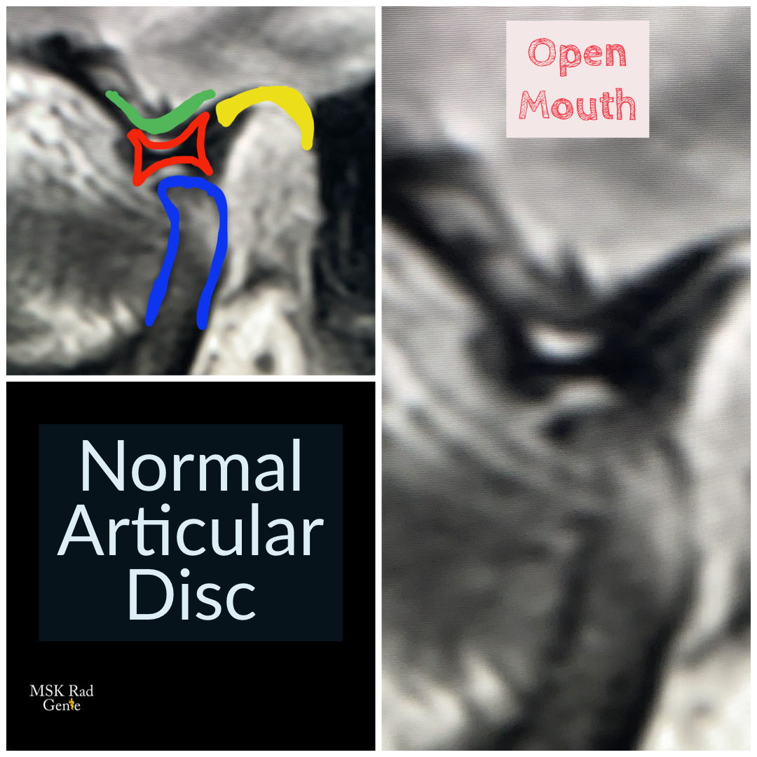
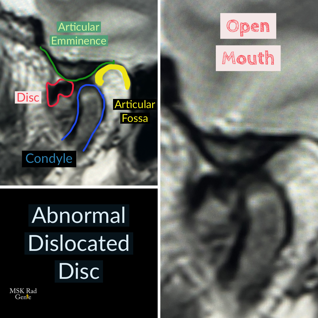
Temporomandibular Joint (TMJ) Disc Dislocation! Dynamic TMJ MRI is mostly used to evaluate the integrity and function of the articular disc. The normal TMJ disc is a biconcave structure that lies between the mandibular condyle head and the articular eminence. It aids in the anterior gliding of the mandibular condyle during open mouth maneuver. With degeneration, the disc may lose it’s normal biconcave configuration and dislocate anterior **The articular disc should always be in top of the mandibular condyle, never anterior or posterior to it** On dynamic MRI, the dislocated disc may either reduce (with an audible “pop”) when the mouth is open, or it may remain dislocated anteriorly. This case shows a dislocated disc that does not reduce upon open mouth maneuver. Clinically, this patients have TMJ pain and inability to fully open the mouth. Many other conditions are evaluated with a TMJ MRI, but disc dislocation is one of the most common pathologies. #mskrad #radiology #radres #radiologia #radtech #radiologystudent #radiologytech #tmjdisorder #jawpain #msk🧞♂️