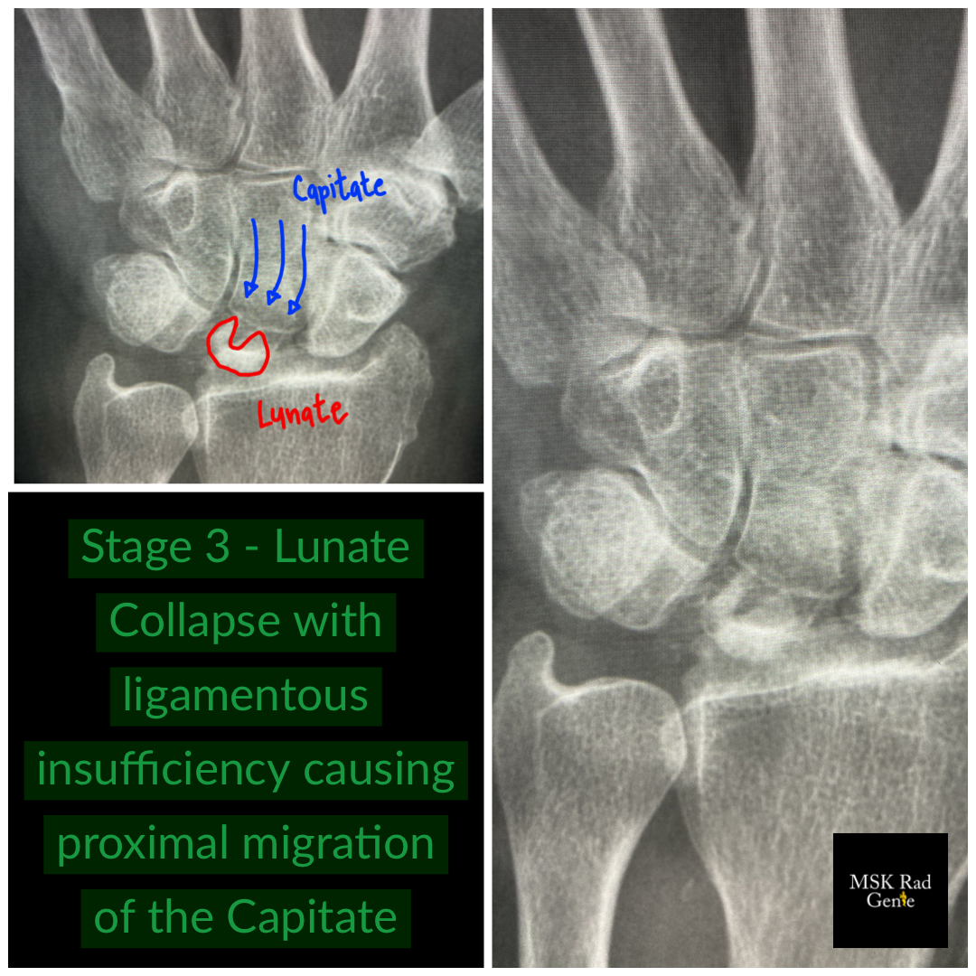

Kienböck’s Disease! Avascular necrosis of the lunate is usually associated to repetitive trauma, although the exact etiology is not precisely known. The lunate has a peculiar and delicate vascular anatomy, which predisposes to AVN when compared to the other carpal bones. The small vessels that feed the lunate run next to the stabilizing ligament – thus any ligamentous injury is usually accompanied to vascular injury. Kienböck is also associated to ulnar negative variance, a common test question. Staging by imaging is simple and requires both radiographic and MRI evaluation. Stage 0 and 1 are only seen on MRI, with simple marrow edema and the treatment is conservative. Stage 2 is seen both on radiograph and MRI and consist of early sclerosis and diffuse marrow edema – treatment is usually surgical during stage 2. Stage 3 and 4 are easily evaluated in radiographs and consist of lunate fragmentation, collapse and involvement of surrounding osseous structures. #mskrad #radres #radiology #radiologia #radiologyeducation #kienbocksdisease #avn #radtech #msk🧞♂️ #mri